Invited Speakers
This is a list of the symposium's invited speakers
Prof. John Barbur, City University London, UK
 | Background:Prof. John Barbur is Professor of Optics & Visual Science and Director of the Applied Vision Research Centre at City University London. More information to be found on his Institute's web page. |
Title: "Assessing the severity of colour vision loss - implications for occupational environments"
Abstract:
Either the absence or abnormal functioning of red / green (RG) or yellow / blue (YB) chromatic mechanisms leads to reduced chromatic sensitivity as well as changes in the perceived colour of objects. This in turn can cause a reduction in the ‘effective’ contrast or conspicuity of objects with potential repercussions on visual performance in visually-demanding working environments.
The use of colour in transport and other working environments has increased significantly in recent years, largely as a result of rapid advances in colour display technologies and lighting systems. Normal trichromatic colour vision, which is rarely enforced, is however often required for employment in such environments. A percentage of subjects with mild congenital RG colour deficiency can carry out suprathreshold colour-related tasks with the same accuracy as normal trichromats and should therefore be classed as safe. In addition to screening for colour deficiency, it has become important to be able to also assess accurately the severity of loss and to establish ‘pass’ limits that do not disadvantage these subjects.
Statistical outcomes based on current methods employed to detect congenital deficiency and to assess loss of chromatic sensitivity will be presented. The relative importance of the factors that cause differences in chromatic sensitivity in normal trichromats and the almost continuous loss of RG chromatic sensitivity in subjects with congenital deficiency will be discussed together with the problems of setting ‘pass’ limits that can be classed as safe without failing unfairly those subjects that can perform as well as normal trichromats. Normal aging and the loss of colour vision in subjects with systemic diseases such as diabetes will also be discussed in relation to colour vision in employment.
Dr. Andreas Bartels, Tübingen
 | Background:Dr. Andreas Bartels is Head of the Research Group for visual perception at the Centre for Integrative Neuroscience, University Tübingen. More information to be found on his Institute's web page.
|
Title: "Colours in the human brain: of movies, the binding problem, constancy, and predictive coding"
Abstract:
Colours are an illusion created by the brain: our colour percept does not represent the light reaching the eye from a given surface, but instead it represents the brain's estimate
of that surface's reflectance. Colours hence appear constant even if the illumination and therefore the reflected light changes - this is referred to as colour constancy.
The neuroscience of colour is hence also the science of conscious perception.
I will review several experiments carried out in our lab that attempt to shed light on neural processing of colour in the human brain: do colours in Mondrians activate the same circuits as colours in movies? Is there a binding problem between colour and motion?
How do memories of object colours influence neural processing?
Where does the brain represent information of the (changing) illuminant and where of (constant) surface reflectance, i.e. colour?
Apart from the above I will briefly introduce key technologies such as brain imaging and multi-variate pattern analysis and the concept of predictive coding.
Prof. Matthias Bleyl, Weissensee School of Art, Berlin
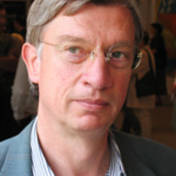 | Background:Prof. Dr. Matthias Bleyl is Professor of General Art History, particularly of the 20th century. More information to be found on his Institute's web page.
|
Title: "Colour - from means of representation to object of representation"
Abstract:
Any painting, depicting real objects in a mimetic sense, uses colours as a means of representation. But, being anyway an abstraction from nature, painting allows a wide range of articulation of the own values colours have to offer. Colour is always bound to the object (local colour), but in painting it has often developed its own life. In painting colour perception always went beyond colour perception in nature, and it is here where the artistic value of a painterly art work is based. Already in figurative painting an expressive use of colour can lead to a considerable shift between local colour of the object and emotional colour perception. In non-objective painting the traditional connection to colour perception in nature is definitively given up, and consequently colour in painting, especially in the second half of 20th century, became itself the object and no longer pure means of representation. A painting of this kind is first of all an object of painterly self-reflection and often an occasion for the reflection on the nature of our perception, not only for the artist but also for the viewer. Its post Enlightenment, informative impact is a specific form of perception, developed in Western democracies and even today rather unwelcome in undemocratic regimes. Some newer positions of the use of paint respectively colour shall illustrate and clarify the artistic capacities colour perception offers to the viewer.
Prof. Justin Broackes, Brown University, Rhode Island, USA
 | Background:Prof. Broackes is Professor of Philosophy at Brown University, Rhode Island. His current work focuses amoung other on the theory of perception and on color and color blindness.More information to be found on his Institute's web page.
|
Title: "Colour blindness and Coloured Filters: What Dalton saw about the attenuation of colour vision"
Abstract:
Dalton believed that his colour blindness (deuteranopia) was due to some kind of blue filtration in the media of the eye: and thought that in candlelight he could pretty well correct many of the errors of his daylight vision. Modern colour research has tended to smile at these suggestions. But for various forms of anomalous trichromacy, mathematical modelling (using shifted response functions for hybrid photopigments) can put a numerical measure on the degree of attenuation in different dimensions of colour variation in viewing a range of coloured surfaces. And for filtration with strong blue and yellow filters (e.g. Kodak Wratten #78 and #86), it can do a similar job. The results can be presented in the form of a Macleod-Boynton diagram: for some purposes a projective transformation of that space, such as a traditional x, y chromaticity diagram is more perspicuous.
The numbers themselves are not to be taken too seriously; but the patterns of similarity between the two sets of phenomena are telling: moderate protanomaly and deuteranomaly are indeed similar to the use of a strong blue filter in reduction of range of RG variation, while leaving the range of BY variation pretty much unchanged. A blue filter is indeed not a bad way for normal trichromats to get an idea of moderate loss of RG variation. What then of Dalton’s no doubt actually dichromatic vision? I shall raise some questions about the difficulties posed by trichromatic behaviour widely reported as occurring in dichromats, and on some of the various factors that might be available to underlie it.
Prof. Axel Buether, Bergische Universität Wuppertal
 | Background:Prof. Buether is Professor of Media Design at the Bergische Universität Wuppertal. More information to be found on his Institute's web page.
|
Title: "Color perception and Memory - The impact of color on our experience and behavior"
Abstract: In the first step of our psychological experiment about 500 participants explored and documented the effects of 13 “psychological primary colors” (Berlin and Kay, 1969) over a period of 6 years. Subsequently more than 1 million images were systematically evaluated.
The aim of the study was to show our everyday knowledge about colors and to find a form of visualization for the complex effects of colors on our experience and behavior.
Our “Color-Memory-Maps” show the complex structure of color memory and its effects on human behavior, which varies with context. Our findings will be presented for the first time to the scientific community at the symposium.
Prof. Bevil R. Conway, Wellesley College, USA
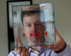 | Background:Bevil Conway is the Sidney R. Knafel Assistant Professor of Neuroscience at Wellesley College and a fellow of the Radcliffe Institute for Advanced Study at Harvard University. More information to be found on his Institute's web page.
|
Title: "Comparing color systems in monkeys and humans"
Abstract:
Patient studies and human neuroimaging implicate specific cortical regions of the ventral visual pathway in the analysis of color. But little is known about the organization of these color-processing regions relative to other functional domains, the causal role of these regions in perception, their connectivity, the precise computations conducted within them, or the underlying neural circuits. The most powerful methods for answering these questions require a primate model. Such a model would be most informative if the processing mechanisms were homologous to those found in humans. How similar then are the cortical mechanisms for color vision in non-human primates and humans? I will discuss recent experiments using psychophysics and neuroimaging that allow a direct comparison of the human and monkey visual system. The work showcases color as a powerful model system for understanding how the brain works, and provides strong evidence that the cortical processing of color is virtually identical between monkeys and humans. Time-permitting, I will discuss preliminary results comparing the cortical responses to auditory pitch (an auditory analogue of color). These findings provide a rare example of a substantial difference in the functional organization of sensory cortex between humans and monkeys.
Prof. David H. Foster, University of Manchester, UK
 | Background:Prof. Dr. David Foster is Professor of Vision Systems at the School of Electrical and Electronic Engineering, The University of Manchester, Manchester M13 9PL, UK More information to be found on his Institute's web page.
|
Title: "Why Colour Constancy Needs More Than Colour"
Abstract: Colour constancy describes the invariance of the perceived colours of surfaces despite changes in the illumination spectrum. It is often explained by simple operations on signals from the cone photoreceptors of the eye. These operations include von Kries scaling by average surface colour (grey-world assumption), scaling by the brightest colour (bright-is-white assumption), and optimizing the gamut of colours. The aim of this work was to test whether such operations can explain colour constancy in a naturally changing environment. Cone signals were calculated from time-lapse hyperspectral radiance images of five outdoor scenes containing mixtures of herbaceous vegetation, woodland, barren land, rock, and rural and urban buildings. Estimates of Shannon’s mutual information between signals were derived across successive intervals of time. Each estimate sets a theoretical upper limit on the number of points in a scene that can be identified by constancy operations. For all five scenes, the number of points declined markedly with increasing time interval, though not always monotonically. The cause was not the change in illumination spectrum as such, but changes in its geometry, for example, the movement of shadows and changes in mutual illumination over the day. It seems that colour signals alone are not enough to identify surfaces in a changing environment. Other signals related to the spatial distribution of colours must also contribute to colour constancy.
Prof. Karl Gegenfurtner, University of Gießen
 | Background:Prof. Dr. Karl Gegenfurtner is Professor of Psychology at the University of Gießen. The emphasis of his current research is on information processing in the visual system.
More information to be found on his Institute's web page. We are very sorry, that due to a conflict in appointments Prof. Gegenfurtner had to cancel his talk at our symposium. He will be awarded the very honorable Wilhelm-Wundt-medal of the German Society for Psychology at the annual meeting in Leipzig on Sept. 19th and 20th, 2016.
|
Prof. Anya Hurlbert, Newcastle University, Newcastle, UK
 | Background:Prof. Dr. Anya Hurlbert is Professor of Visual Neuroscience at the Newcastle University, Newcastle, UK. The emphasis of her current research focuses on human visual perception. More information to be found on her Institute's web page.
|
Title: "Seeing (and Feeling) the Light"
Abstract:
People see objects as having colours – red strawberries, yellow lemons, blue cornflowers – because of interactions between light, surfaces and the sensors in the eyes, which initiate further processing in multiple brain areas. One of the fundamental properties of this perceptual processing is colour constancy, the phenomenon by which object colour remains constant despite changes in the illumination spectrum – the yellow lemon stays yellow whether illuminated by tungsten light or morning daylight. Its colour may therefore serve as a reliable indicator of its identity and edibility.
Yet colour constancy in not perfect. In everyday experience, people not only see alterations in colour appearance of objects but also changes in the colour of the light. In this talk, I will explore the notion of object colour constancy as perceptual insensitivity to changes in the illumination spectrum over time. I will describe behavioural measurements of illumination discrimination made using real objects and spectrally tuneable multi-channel LED light sources. Results from these experiments suggest that colour constancy is optimised for natural illuminations, and that people are least sensitive to changes in illumination along the cooler, bluer end of the daylight range. In other experiments examining the non-visual effects of spectral changes in illumination, we find that bluer lights also have greater capacity to suppress sleepiness, yet are less preferred than warmer lights. Thus, people both see and feel the light. Given the continuing technological innovations in artificial lighting, it is all the more important to understand how these responses to light affect people’s perception of object colour.
Prof. Gerald Jacobs, University of California, Santa Barbara, USA
 | Background:Prof. Dr. Gerald Jacobs is Research Professor of Psychology and Brain Sciences at the University of California, Santa Barbara. Research in his laboratory is broadly centered on issues having to do with the biology of mammalian vision. More information to be found on his Institute's web page.
|
Title: "A Comparative Look at Photopigments and Color Vision"
Abstract:
The widespread adoption of molecular biological techniques, in conjunction with an understanding of the linkages between the structure of opsin genes and their photopigment products, has yielded predictions about the spectral properties of photopigments found in a large number of invertebrate and vertebrate species. The full complement of photopigments present in any animal sets limits on what it may be able to see, while within those boundaries opening a broad range of visual possibilities. Given that, it is not surprising that these measurements are often—in fact, almost routinely—used as grounds for drawing inferences about the details of an animal’s color vision. One of the most common inferences is about its dimensionality, i. e., is the animal likely to be a dichromat, a trichromat, a tetrachromat, or perhaps even something beyond that? At the root, such linkages are based on our understanding of human color vision. I’ll describe cases that illustrate how such conclusions have been extended—sometimes with corroborating evidence, but most often without—to further claims about color vision in nonhuman species. Among other things, the literature reveals numerous examples where the standard human model fails to capture the ways in which different species have evolved a capacity to exploit signals gathered from the presence of multiple types of photopigment.
Dr. Gabriele Jordan, Newcastle University, Newcastle, UK
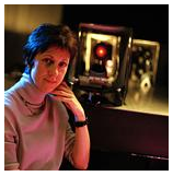 | Background:Dr. Gabriele Jordan is senior lecturer at the Institute of Neuroscience at Newcastle University, Newcastle, UK. The emphasis of her current research focuses on the inter-individual variabilities seen in X-linked (red/green)colour vision of both colour-normal and colour-deficient observers. More information to be found on her Institute's web page.
|
Title: Discriminating colours in tetrachromatic space
Abstract:
Colour vision is often taken for granted and many people assume that our perceptual worlds are equally colourful. However, this is not the case since marked inter-individual differences exist both within the colour-normal population as well as within the sub-group of individuals diagnosed as colour deficient. The perceptual differences are caused primarily by the variability of two X-linked genes coding for the middle- (M) and long-wave sensitive (L) cone photopigments in the retina.
About 12% of women are carriers of anomalous trichromacy caused by a red/green hybrid gene giving rise to a cone photopigment with a spectral sensitivity somewhere between those of the normal M and L cone photopigments. Random X-chromosome inactivation ensures that the retinal mosaic of such a carrier will contain four rather than three types of cone and people have speculated whether individuals with such four-cone retinae could enjoy four-dimensional colour vision.
We have been able to lend strong support for this hypothesis (Jordan, Deeb, Bosten & Mollon, 2010), but overall our results suggest that tetrachromacy is not afforded automatically to those with four types of retinal cone. I will describe psychophysical tests that have led to our conclusion and will outline the challenges of measuring tetrachromatic colour vision
Prof. Jan Kremers, University Hospital Erlangen
 | Background:Prof. Dr. Jan Kremers is Professor of experimental Ophthalmology at the University of Erlangen-Nuremberg. More information to be found on his Institute's web page.
|
Title: "Electrophysiological correlates of cone-opponent processing in the human retina"
Abstract:
The electroretingram (ERG) is often used in a clinical environment to assess the functional integrity of the retina. The significance for studying retino-geniculate pathways has been limited. The work of Jerry Jacobs and colleagues have demonstrated that an heterochromatic flicker photometric (HFP) paradigm yielded data that resemble those obtained in psychophysical experiments studying the magnocellular (luminance) pathway.
We have demonstrated over the last few years that ERG responses to combined sinusoidal modulation of luminance and chromatic modulation reflect luminance activity at high temporal frequencies (>30 Hz; confirming the results of Jacobs’ studies) and red-green chromatic activity at intermediate frequencies (about 12 Hz). This has been demonstrated by multiple different paradigms. From these data we conclude that the ERGs are governed by two fundamentally different mechanisms, which are probably related to retino-geniculate pathways with relevance for vision.
We propose that one of the reason that this correlation with the activity of retino-geniculate pathways was not noticed before is that mainly flashed luminance stimuli were used in previous studies. In subsequent studies, we demonstrated that ERG responses to instantaneous changes in excitation of only the L-cones or only the M-cones have opposite properties: The responses to L-cone increments (“L-On”) and to M-cone decrements (“M-Off”) resemble each other. In addition, ERGs to L-cone decrements (“L-Off”) and M-cone increments (“M-On”) are very similar. These data indicate, that the ERGs are to a large extent determined by L/M cone opponent processes. In a psychophysical experiment, we further demonstrated that L-On flashed stimuli are perceived as brightness increases, whereas M-On stimuli are perceived as brightness decreases. This is another demonstration of the close correlation between ERG responses and psychophysical data that rely on selective activities of retino-geniculate pathways. These data also indicate a new dimension in the correlation between the two because this is the first time that a psychophysical experiment was designed based on ERG data instead of the opposite.
We further demonstrate that the two ERG mechanisms have different spatial properties: The amplitudes of luminance reflecting ERGs are monotonously correlated with stimulus sizes. Their phases do not depend strongly on stimulus size. In contrast, ERGs that reflect L/M cone opponency do neither change in amplitude nor in phase when the stimulus size exceeds a certain area. For smaller stimuli, both amplitude and phase depend strongly on stimulus area.
Finally, we found evidence that the two mechanisms may be differently affected by retinal disorders. Particularly, patients with Duchenne’s muscular dystrophy show distinct defects in the ERGs reflecting luminance and L/M-cone opponent mechanisms.
In conclusion, ERG responses are governed by two fundamentally different mechanisms, which are probably related to luminance and L/M opponent retino-geniculate pathways with relevance for vision. The ERGs can be used to study properties of these pathways with non-invasive electrophysiological techniques in human observers.
Prof. Ichiro Kuriki, Tohoku University, Japan
 | Background:Prof. Ichiro Kuriki is Associate Professor at the Research Institute for Electrical Communication, Tohoku University. More information to be found on his Institute's web page.
|
Title: "Cortical response to categorical color differences in prelinguistic infants"
Abstract:
We often use categorical color expression in words when communicating about color. To examine whether the color category is formed by the acquisition of language, we measured brain activity in prelinguistic infants (5-7 months old). We presented two types of color pairs: color alternations within a color category or between two color categories. The colors were selected to span the border of green and blue categories with equal color difference steps. Infant’s categorical border was confirmed by a behavioral experiment using habituation method; the infants preferred to look a test color only when it is chosen from a novel category. The infants’ cortical responses were measured by near infrared spectroscopy (NIRS) in the occipito-temporal area and it showed significant signal changes when the color altered across color categories. Such a difference in brain responses, in relation to the color category, was not found in occipital area responses. The occipito-temporal response was also found in adults, but lateralization was not observed in both infants and adults. The lack of laterality and the presence in prelingual infants implies that the NIRS responses in the occipito-temporal region may be corresponding to the brain activity is possibly related to non-verbal color categories. Considering the analogy to the NIRS responses in relation to phonetic categories in prelingual infants, it is possible to infer that categorization of visual inputs has already started before the acquisition of relevant words and the category would be modified by language, later on.
Prof. Barry Lee, MPI Göttingen / SUNY, New York
 | Background:Prof. Dr. Barry Lee is Professor of Vision Science emeritus at the State University of New York. More information to be found on his current page.
|
Title: "Segregated transmission of achromatic and chromatic signals in the primate visual pathways"
Abstract:
The primate visual pathway contains three main cell pathways, the magnocellular (MC), parvocellular (PC) and koniocellular (KC) systems. The MC pathway underlies the luminosity function, whereas the PC and KC pathways respond well to stimuli related to the red-green or blue-yellow dimensions of colour experience. It is often held, however, that the PC pathway does double duty and plays a major role in achromatic spatial vision. I shall review assumptions of this model, and argue instead that the PC pathway is a most unusual ganglion cell class, highly specialized for transmission of an |L-M| opponent signal. For example, the midget morphology does not bear any relation to center size; one reason for this is that the point spread function is much larger than cone or dendritic tree diameter. It is much more plausible that midget morphology is concerned with providing a cone-specific signal to inner retina. I shall then describe some experiments supporting the idea that achromatic, red-green and blue yellow information is strictly separated in the MC, PC and KC pathways. For example, physiological and psychophysical experiments with gratings containing luminance and chromatic components of different spatial frequencies demonstrate strict segregation of signals. Lastly, recent physiological data show the MC pathway well adapted to transmitting fine spatial detail; much energy is concentrated in their responses to the fine detail in spatial patterns. These arguments suggest the double duty model is not tenable, but are consistent with, for example, efficient coding arguments of MacLeod and coworkers.
Prof. John Mollon, Cambridge University, UK
 | Background:Prof. John Mollon is Professor of Visual Neuroscience at the University of Cambridge, and College Lecturer and Assistant Director of Studies in Psychology, Gonville and Caius College, Cambridge. More information to be found on his Institute's web page.
|
Title: "Seeing colours as different"
Abstract:
In a powerful paper of 1949 (Revue d'Optique, 28, 262-278), Yves Le Grand re-analysed the colour-discrimination data that MacAdam had obtained at different positions in colour space. Estimating the spectral sensitivities of the retinal cones from the matches of colour-blind observers, Le Grand calculated how the cone signals were modulated as chromaticity was varied along lines of different direction through a given point in colour space. He concluded that colour discrimination depends on two independent signals, which correspond to S/(L+M) and L/(L+M), where L, M, S are the excitations of the long-, middle- and short-wavelength cones respectively. In the case of the first signal, Le Grand suggested, thresholds increase with the log of S/(L+M), whereas L/(L+M) discrimination is minimal at the L/(L+M) value of the adapting white.
There prove to be many complications to Le Grand’s attractive account. Danilova and I have found that (i) Discrimination along a horizontal axis in the MacLeod-Boynton diagram depends on the level of S-excitation; (ii) Thresholds are not minimal at a fixed value of L:M, corresponding to the L:M ratio at the adapting chromaticity, but instead are minimal at the boundary between reddish and greenish hues ¬– a boundary that corresponds closely to the ‘caerulean line’ (the locus of natural illuminants); and (iii), Saturation thresholds are systematically higher than hue thresholds, even when all that differs is the phase in which the underlying ‘cardinal’ signals are combined.
One further issue of colour discrimination has enjoyed little attention: What is the mechanism that allows us to compare colours with precision when they lie far apart in the visual field, even in different hemifields?
Prof. Jay Neitz, University of Washington, Seattle, USA
 | Background:Prof. Jay Neitz is the Bishop endowed Professor of Ophthalmology at the University of Washington, Seattle. More information to be found on his Institute's web page.
|
Title: "How the world became colored: the evolution of conscious color perception in primates"
Abstract:
Nearly 40 years ago, in his book “Human Color Vision,” Boynton wrote “The chromatic code of the visual nervous system is incomplete and difficult to interpret;” however, a series of recent discoveries are providing the missing puzzle pieces needed to complete a picture of the diversity of ganglion cell types involved in primate color vision and their varied functions. Boynton proposed a model that, in its simplest form, has just two color channels, red-green (RG) comparing L vs. M cones and blue-yellow (BY) comparing S-cones to the other two types. Neurobiological explanations of color vision have focused on the two ganglion cell types that most closely correspond to the channels Boynton described, small bistratified for BY and midget ganglion cells with L vs. M opponency for RG. However, in contrast to this simple idea, recent evidence suggests that there are many ganglion cell types and subtypes in the primate retina that carry color information. These have appeared at vastly different times over the history of the evolution of vertebrates, they project to several different places in the brain and serve a variety purposes. Many appeared before mammals evolved; including color coded ganglion cells involved in the modulation of sleep and mood, guidance of movements, detection and object segmentation. In contrast, based on recent results we suggest that four specific types of chromatically coded ganglion cells evolved exclusively in primates for the uniquely anthropoid function of assigning blue, yellow, red and green colors to objects for use in identification and classification.
Prof. Karl Schawelka, Bauhaus University of Weimar
 | Background:Prof. em. Dr. Karl Schawelka is Professor of Art History at the Bauhaus University of Weimar. His scientific focus is on color and perception, Theory of Art, Contemporary Art and Art in public spaces. More information to be found on his Institute's web page.
|
Title: "The Colours of Paradise and its Discontents“
Abstract:
Paradise is full of saturated and strong colours while the realm of the dead is shadowy and pallid. The locus amoenus or the dreamland arcadia of the ancients or popular pictures in calendars have in common that they show a welcoming and colourful place where it is good to settle and procreate. Multicoloured paradise pleases the senses. It is related to sex.
However, this cannot be the whole story. We often restrain from motley compilations since excess of colour can be unpleasant. There must be at least one opponent process. How do we have to understand this process, its logic and the reason for its existence?
Of course theoreticians have since long noticed that there exists some sort of chromophobia, to use David Batchelor’s term. Usually they blame social rules for it. I am not convinced that these are sufficient to explain the restrained colourfulness we generally show in our actual environments. When we are fed up by an exuberance of colours it’s a spontaneous feeling and we don’t act out of social fear. A temporal factor has to be considered. But not only the balance shifts, the role of the colours - that what they stand for - also changes.
Paradise is attractive when we are safe and able to enjoy. When we must meet urgent tasks, the adequacy of a colour to its functional role becomes imperative. They should be easily distinguishable and semantic and pragmatic distinctions play a greater role.
Prof. Arthur G. Shapiro, American University, Washington DC
 | Background:Prof. Arthur G. Shapiro is Department Chair for Computer Science and for the Department of Psychology. He specializes in the areas of visual perception and cognitive neuroscience and is best known for creating a series of visual phenomena ("illusions") that have arisen from this research. More information to be found on his Institute's web page.
|
Title: Multiple spatial systems for color vision
Abstract:
Color cells in the visual cortex can be divided into several classes based on their spatial characteristics (Conway, 2001; Johnson, Hawken & Shapley 2001, 2008; Conway and Livingstone, 2006; Solomon and Lennie, 2007); most models of perception, however, do not consider the possibility that there are multiple spatial channels for color vision. Here I will present phenomenological and psychophysical evidence for segregating color vision based on two different types of spatial information: 1. A separation between color vision and color contrast vision; and 2. A separation between low spatial frequency and high spatial frequency color vision. As evidence for a separation of type 1, I will discuss explorations with the Contrast Asynchrony paradigm (Shapiro, 2008). The main idea is that a colored disk surrounded by a colored annulus creates separable dimensions for the color of the disk and the color-contrast between the disk and the surround. The separation suggests advantages for representing color in a color plane with rectified contrast on one axis and color (or luminance) along the other axis. For evidence of 2, I will demonstrate that many color and brightness phenomena can be explained simply by removing low spatial frequency content (see Shapiro and Lu 2011). I further demonstrate that under many conditions, low spatial frequency color information corresponds to changes in illumination, whereas high spatial frequency color information remains invariant to changes in illumination. I will demonstrate some limitations to these divisions and show how these limitations can be overcome by adaptive weighting of spatial information.
Prof. Andrew Stockman, UCL, London
 | Background:Prof. Andrew Stockman is the Steers Professor of Investigative Eye Research at the Institute of Ophthalmology, University College London. More information to be found on his Institute's web page. and the web page of CVRL - Color & Vision Research Laboratory.
|
Title: "Distorted insights: from hue anomalies to colour mechanisms"
Abstract:
Observers viewed slowly-on and slowly-off, red-green-chromatic, flickering sawtooth-waveforms. Between about 5 and 13 Hz the mean hue of the sawtooth waveforms shifted in the hue direction of their shallower slope, a result consistent with the hue-change being limited by a “slew-rate” process that is better able to follow slower or more prolonged changes in the waveforms and therefore shifts the mean hue in that direction. Importantly, this process follows substantial temporal filtering so that the phenomenon depends mainly on the 1st and 2nd harmonics of the waveforms.
We next presented just the 1st and 2nd harmonics of the sawtooth stimuli and investigated the dependence of the time-varying (AC) and the mean (DC) hue-appearances on the phase of the 2nd harmonic. Between 0.67 and 4 Hz, the hue-appearance is dominated by an AC hue-variation that depends on the amplitudes of the composite waveforms rather than on their slopes. At these frequencies, the results are consistent with peak sensors that register the maximum excursions of the waveforms in the red and green hue directions. At frequencies between 5 and 13 Hz, where the slew-rate limits the chromatic signal, the hue-appearance is more complex and is seen as an AC hue-variation around a mean DC hue that is shifted away from the true mean. At these higher frequencies, the results are also consistent with a peak sensor, but the hues are affected by a slew-rate limited signal that regulates the AC signal.
Prof. Peter Tse, Dartmouth College, USA
 | Background:Prof. Peter Tse is Professor of Psychology and Brain Sciences at Dartmouth College. More information to be found on his Institute's web page.
|
Title: "The neural basis of color filling-in and its attentional modulation"
Abstract:
The traditional view of filling-in has been that a central color is replaced by a surrounding color once borders have faded, as in the Troxler illusion. We find instead that colors mix from inside to outside and outside to inside. We find neuronal evidence for this mixing as early as V1. I will also show data that suggest that attention can influence the process of filling in by specifying which boundaries the bottom-up processes will be computed within.
PD Dr. Gregor Volberg, University of Regensburg
 | Background:PD Dr. Volberg is a senior lecturer in the department of Psychology at the University of Regensburg. His research focus is on neural connectivity in visual attention and perception, obtained from oscillatory brain activity in the EEG. In a DFG-funded project he investigates the neuro-cognitive mechanisms of grapheme-color synesthesia. More information to be found on his Institute's web page.
|
Title: "Seeing colors in achromatic stimuli: Grapheme-color synesthesia"
Abstract:
Synesthesia is a rare perceptual phenomenon where unimodal stimuli induce concurrent sensations in a sensory modality that was not objectively stimulated. One of the most investigated forms is grapheme-color synesthesia where written language symbols like numbers or letters induce concurrent sensations of color.
A key question on grapheme-color synesthesia is at which level of processing the color co-activation occurs. One position is that the color co-activation is a perceptual process that emerges during form processing of the grapheme. The other extreme is marked by the idea that the color co-activation roots in memory rather than perception, and that color is part of the object knowledge for specific graphemes.
I will argue that electroencephalography (EEG) with its fine temporal resolution is an appropriate method for investigating the neural dynamics underlying grapheme-color synesthesia. Our EEG experiments revealed increased activity in color- and grapheme-processing brain areas in synesthetes for color-inducing compared to non-color-inducing graphemes, even under passive viewing conditions where no processing for meaning was required. Color-inducing compared to non-color inducing graphemes also produced higher phase-locking between EEG oscillations (i.e. an increased functional connectivity) between frontal and posterior sites. Our results suggest that grapheme-color synesthesia is perceptual in nature, and that the color co-activation relies on inter-areal neural communication.
Prof. Dr. Christoph Wagner, University of Regensburg
 | Background:Prof. Dr. Christoph Wagner is Professor of Art History at the University of Regensburg. His fields of interest are, among others, paintings from 1500 until today, and Art Theory and Art in Practice of the European avant-garde. More information to be found on his Institute's web page.
|
Title: ""Interaction of Color" – Concepts of Seeing Colors in Modern Art."
Abstract:
No epoch of art history has shown a greater fascination of the elementary phenomena of color perception than the modern age: corresponding to scientific theories of psychology and neurology various concepts of seeing colors have developed as the basic principles of art. Chevreul’s and Helmholtz’s pioneering analyses of the incongruity of optical, physiological and aesthetic dimensions have tread this path already in the 19th century. In this context Helmholtz established the notion of painting as a colored “translation”: “In order to imitate nature, color needs to be depicted in a different way within a painting“ (Chevreul). It is “the contradiction between physical facts and psychological effect” which causes new ideas of “interaction of color”. For Josef Albers this meant nothing less than the “origin of art”. Artists of the second half of the century have developed the vision of color as a pure visual entity, that is freed from material pigment, in manifold ways. My talk will focus on the question how new strategies of aesthetic effects and concepts of seeing colors influence the artists’ attempts to transform the use of color into an optical, often virtual constant that loosens its bond to classical coloring.
Prof. Michael A. Webster, University of Nevada, Reno, USA
 | Background:Prof. Michael Webster is Foundation Professor of Cognitive and Brain Sciences at the Department of Psychology at the University of Nevada, Reno. More information to be found on his Institute's web page.
|
Title: "Blue and yellow in the world, the brain, and the dress"
Abstract:
Blue and yellow hold a special place in color theory as one of the principal dimensions underlying color appearance. However the basis for their special nature as color percepts remains a mystery. This talk will examine the relationship between blue and yellow in the environment and the brain, and how this relationship is shaped by visual adaptation. Blue and yellow are often conceived as opponent poles of a single dimension, yet there are a number of asymmetries between them, and like other hues they vary independently from one observer to the next. We have also discovered a striking difference in how they are experienced, such that blues are more likely than a complementary yellow to be appear ambiguous or as a shade of gray. This asymmetry may underlie a number of phenomena of color and material perception including how people perceive the colors in “the blue and black (or yellow and white) dress,” and may reflect a bias to attribute blue to the lighting but yellow to the object.
Prof. John S. Werner, University of California, Davis, USA
 | Background:Prof. John S. Werner is Distinguished Professor in the Department of Ophthalmology and Vision Science at the UC Davis School of Medicine. More information to be found on his Institute's web page.
|
Title: "Phenomena of Color and the Quest for Mechanisms"
Abstract:
Human vision begins with the absorption of light by pigments within photoreceptors that transform energy into electrochemical signals. These signals are further processed by a cascade of cellular interactions in the retina and higher areas of the brain that ultimately result in our ability to perceive hue, saturation and brightness. The initial stages of neural processing are well understood such as the ability of normal individuals to match any wavelength of light with a combination of three other wavelengths, a capacity called trichromacy. This is due to the presence of three types of cone photopigments that differ in their peak absorption at either short (S), middle (M), or long (L) wavelengths. Within the retina, cone signals are transformed into ratios (e.g., S vs. M+L and L vs. M) which form the basis of wavelength discrimination. This cone opponency does not, however, explain our perception of color, experiences that Hering described in terms of red-green, yellow-blue, black-white opponent pairs. The latter are sensations that oppose each other; that is, the sensations of red and green, and blue and yellow, cancel each other. These opponent processes also operate over space and time to give rise to numerous phenomena of color contrast. Indeed, many colors (e.g., dark colors such as brown or black) are experienced only with preceding or surrounding contrast. Although many phenomena of color perception are treated popularly as illusions, they may point to more fundamental processes that adjust color coding to optimize detection and discrimination of colors, segmentation of figure and ground, while maintaining color constancy. The neural substrate for these processes is not well understood, or agreed upon. Indeed, some linguistic-anthropological studies even question whether Hering’s aforementioned hue terms are necessary and sufficient to describe the color experiences of all cultures. This skepticism is buttressed by findings from both single cells in the cortex and perceptual studies indicating that color channels can be tuned to many different directions in color space, not just to red-green and yellow-blue axes.
Prof. Sophie Wuerger, University of Liverpool, UK
 | Background:Prof. Sophie Wuerger is Professor of Psychological Sciences at the Institute of Psychology Health and Society, at the University of Liverpool. Her research focuses on age and color perception. More information to be found on his Institute's web page.
|

