x-ray crystallography
X-ray crystallography and cryo-electron microscopy (cryo-EM) are complementary methods in the field of structural biology. Depending on the specific research question, our group conducts x-ray diffraction experiments when the protein of interest can be crystallized in the desired state. We consider two objectives when choosing between the two methods:
1. Sample Size: Cryo-EM is particularly well-suited for the analysis of large macromolecular complexes and flexible proteins across a wide range of molecular masses. In contrast, x-ray crystallography is more suitable for smaller protein structures.
2. Resolution: Cryo-EM has made significant advancements in recent years and is capable of achieving near-atomic resolutions, typically ranging up to 1.5 Å. X-ray crystallography, meanwhile, generally provides higher-resolution data, allowing for more detailed structural information.
By considering these factors, we can select the most appropriate method to obtain the desired structural insights for a given protein or complex.
Equipment:
Pipeting robotics: mosquito LCP for membrane protein crystallization enables accurate, reproducible automation for liquid cubic phase (LCP) setups:
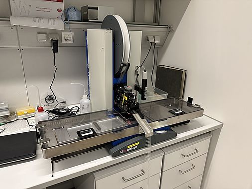
Crystallization incubators allowing for crystal formation at constant temperatures:

Crystal handling bench equipped with advanced light microscopes and imaging system:

Data collection is performed remotely at SLS PX I and PX III:

Christine Ziegler - 08.09.2023 18:18 