Scanning-AUGER Spectroscopy – Sc-AUGER
General Description
The Scanning AUGER Spectrometer comprises a scanning electron microscope (SEM) and an energy analyzer electron energy spectrometry. The SEM offers a lateral resolution of about 200nm. The energy analyzer consists of a cylindrical mirror analyzer and a multichannel detector. Due to the physical origin of the analyzed electrons (the Auger photoemission process) the Sc-AUGER is more sensitive to lighter elements compared to XPS. One of the main advantages of Sc-AUGER is the Chemical Mapping Mode which reveals the chemical composition of the sample surface as well as chemical spectroscopy in a predefined area on the sample with a resolution given by the SEM, see Measurement Modes.
Measurement Modes
- SEM Scan Mode
This microscopy mode is used to display the sample surface. The maximum resolution is about 200nm. - Spectrometry
The sample surface is analyzed with respect to its chemical composition in a region predefined within the Scan Mode. - Chemical Mapping Mode
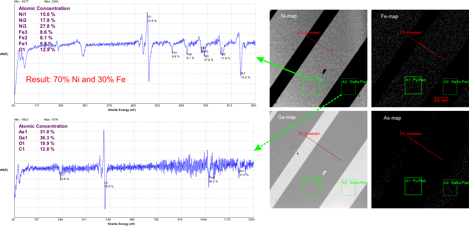
The chemical composition of the sample surface is displayed as map on the sample surface. The lateral and energetical step size are parameters to be defined. An example of such a chemical map is shown in Fig. 1. - Depth Profile
During a continuous Ar-ion sputtering process which removes the sample layer-by-layer the element-specific profile across the sample is recorded.
Example: NiFe stripes on a GaAs-substrate
Sample Structure
The dimension of the samples to be investigated should be from 2 by 1 mm to maximum of about 2 inch or 51mm.
Margit Adler - 04.03.2021 11:12 