Research
Our research has revealed that the immune system has adapted the evolutionary conserved signalling pathway of the Epidermal Growth Factor Receptor (EGFR).
For instance, we found that the EGF-like growth factor Amphiregulin is a cytokine expressed by leukocytes, contributing to resistance to helminth infection. (Zaiss et al. Science 2006). Recently, we then revealed that Amphiregulin is achieving this by enabling the formation of hetero-complexes / signalling clusters between the EGFR and IL-33R on the surface of Th2 cells. These hetero-complex/clusters enable Th2-cells to express IL-13 upon exposure to IL-33. In this way, at the site of infection, Th2-cells contribute to worm expulsion in an antigen-independent way. (Minutti et al. Immunity 2017).
Furthermore, we discovered that a crosstalk between the EGFR and TGFβ controls local immune responses. Based on a “biased agonism” of the EGFR, the low-affinity EGFR ligand Amphiregulin induces the local activation of TGFβ (Minutti et al., Immunity 2019). In this way, Amphiregulin enhances the suppressive capacity of regulatory T-cells, thereby critically contributing to the resolution of inflammation and tissue homeostasis. (Zaiss et al. Immunity 2013). TGFβ is expressed and stored in tissues in form of a latent complex. As a low-affinity EGFR ligand, Amphiregulin induces a tonic sustained signal which activates integrin-αV complexes on target cells and thus induces the local release of bio-active TGFβ. In this way, Amphiregulin contributes to local immune suppression and the differentiation of tissue residential stem cells, such as pericytes, and thus to tissue repair. (Minutti et al. Immunity 2019).
This rather unexpected concept of local immune regulation challenges and suggests the re-evaluation of several of our present perceptions with regard to inflammation, wound repair and tissue homeostasis; and, thus, also their implications for the development of tissue fibrosis, cancer and auto-immune diseases.
We are further exploring the fundamental implications of this novel avenue of research and apply our findings for the development of a sophisticated therapeutic research programme in order to translate this knowledge to the benefit of patients.
Currently, we are developing research projects on several different levels:
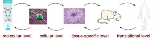
On a molecular level
Cytokine hetero-complexes as a scaffold for cytokine signalling:
We have shown that the EGFR can form hetero-complexes with other cytokine receptors (Minutti et al. Immunity 2017). The formation of hetero-complexes between cytokine receptors is an enigmatic process. We are currently following the assumption that the transient activation of the EGFR induces distinct cluster formations of activated receptors on the cell surface. These clusters are then directed to intra-cellular signalling complexes, which we assume to be actin-anchored. Only in these signalling-competent clusters will then a cross-talk between the different signal transduction pathways be initiated. We are using microscopy-based approaches to resolve these questions.
EGFR signalling in tumour development and cancer therapy:
Several different types of tumours over-express either a wt or mutated form of the EGFR. These types of tumours are often hard to treat, show a “lymphocyte exclusion” phenotype and poorly respond to check-point inhibitor treatment. We propose that similar to Amphiregulin-mediated EGFR activation, also the EGFR complexes on these tumours induce a sustained activation of the PLCγ signalling pathway and thus the local activation of TGFβ. For a review of this concept please see: Kapoor & Zaiss (2021) Biomedicines 27:52.
In order to test our hypothesis, we established together with the MRC Mouse Genetics Research Institute in Harwell a mouse strain with a point mutation in the EGFR (EGFR-Y992F). In this mouse strain, the main selective link between the EGFR and the PLCγ signalling pathway is disrupted. Thus, in this mouse strain the overexpression of the EGFR cannot lead to the local actiation of TGFβ any longer. In mouse models of spontaneous tumours, which are well established to overexpress the EGFR, we will now determine whether the disruption of the link between the EGFR and the PLCγ signalling pathway enhances the immune systems capacity to clear newly developing tumours spontaneously or whether it may enhance the effciacy of anti-tumour treatments.
This set of experiments will demonstrate whether by targeting the EGFR in a clinical setting one influences the local tumour micro-environment and, thus, whether we could use clinically approved EGFR inhibitors as a means to enhance the efficacy of tumour immune-therapy in several different types of cancer.
On a cellular level
To achieve coordination of immune responses, for instance during infections, the EGFR influences the functionality of leukocytes, such as T-cells, during different phases of an immune response:
During the initiation phase:
Using single cell sequencing and TCR cloning, we found that high-affinity TCR ligands induce the expression of HB-EGF, which enhances the survival rate of primed naïve CD4 T-cells and prevents TGFβ-induced Th-17 differentiation of these cells. Thus, EGFR mediated signalling leads to the expansion of high-affinity TCR clones and skews pathogen-specific immune responses towards a Th1 phenotype, thereby preventing auto-immune diseases.
During the effector phase:
Using an adoptive T-cell transfer system into MHC-II-deficient mice, we have been dissecting the mechanisms by which CD4 T-cells use EGFR expression to sense the local state of inflammation (Minutti et al. Immunity 2017). We found that the EGFR forms hetero-complexes with IL-1R related transmembrane receptors (TIR). In this way, T cells can function in an antigen-independent way at the site of infection.
During the resolution phase:
Employing a passive transfer models, we are determining whether HB-EGF expression enables local effector CD4 T-cells to escape TGFβ-mediated immune suppression and thus to maintain local inflammation, for instance, in inflamed arthritic joints
On a tissue specific level
Combing experimental induced inflammation with a intravital microscopy-based approach in reporter mice, we are determining how local inflammation uses the EGFR / TGFβ crosstalk to impose a dynamic, spatio-temporal activity pattern of TGFβ that coordinates pathogen clearance with wound repair.
Using infection models, we are testing whether that at the site of infection, monocyte-derived HB-EGF expression prevails and suppresses TGFβ function and thus sustains efficient local immune response; while in the periphery, Amphiregulin prevails and induces TGFβ activation and thus the resolution of local inflammation; thereby facilitating wound repair by inducing the differentiation of tissue stem cells, as well as of myofibroblasts.
Via an enhanced collagen deposition, this enhanced myofibroblast differentiation contributes to wound healing, the encapsulation of the infection site – but can also leads to tissue fibrosis and loss of organ/lung function.
On a pre-clinical / translational level:
We have been developing neutralizing antibodies and peptide-based inhibitors, which can specifically block the activity of either Amphiregulin or HB-EGF.
The Amphiregulin specific inhibitor prevents and reverts lung fibrosis in a chronic lung inflammation model (house dust mite extract) and a bleomycin model. We intend to develop these inhibitors further for the therapeutic application in lung transplant patients and in patients suffering from Idiopathic Pulmonary Fibrosis (IPF).
Also, we are testing the HB-EGF inhibitor in mouse models for its therapeutic efficacy, i.e. in treatment auto-immune diseases, such as rheumatoid arthritis. HB-EGF enhances the survival of T-effector cells and renders them resistant to regulatory T-cell and TGFβ mediated immune suppression. We predict that within inflamed joints HB-EGF specific inhibitors could mimic a situation observed following TNFα inhibitor treatment, which has been shown to enhance the suppressive capacity of regulatory T-cells in inflamed joints. Thus, HB-EGF specific inhibitors could potentially be utilized to supplement or substitute TNFα inhibitor treatments in their clinical use.
Dietmar Zaiß - 07.02.2022 22:53 