Research
Sensors & Actuators for Animal Cells

Our lab is focused on interfacing animal cells (2D monolayers or 3D aggregates) with physical transducers (e.g. planar electrodes, piezoelectric devices, optical waveguides) to monitor the cell response during toxicological or pharmacological assays non-invasively and in real-time. The non-invasive nature of the measurement provides the dynamics of the cell response as another level of biological insight. It is the longterm goal of our research to replace classical concentration-analysis by effect-analysis on biological organisms in general, and animal cells in particular. To increase the information content we combine individual transducer principles in one setup and include invasive actuators (electroporation, electrofusion, electrodeformation) to expand the scope of label-free analysis.
Impedimetric Whole-Cell Biosensors - Goldfilm Electrodes
Impedimetric Whole-Cell Biosensors
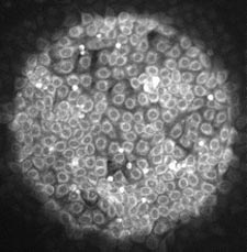
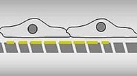
Impedimetric monitoring of adherent animal cells relies on cell-covered goldfilm electrodes that serve as growth substrate and measuring electrode at a time. When the cells attach and spread on the electrode surface, the dielectric cell bodies increase the electrical impedance of the electrode, as the current is now forced to flow around or through the cells. The exact current pathway can be experimentally controlled by the AC frequency of the driving voltage that is used in the experiment.
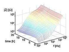
The sketch below illustrates the principle of the measurement with a micrograph of a cell-covered electrode. The figure on the right demonstrates the richness in information when the complex impedance Z is measured as a function of frequency during attachment and spreading of cells upon the electrodes.
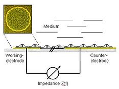
For most applications current flow around the cells is of of greater interest as the measured impedance holds information on cell-cell and cell-substrate contacts. Whenever the cells change their shape in response to a biological, chemical or physical stimulus, this cell repsonse is mirrored in a corresponding change in impedance. Thus, impedance readings basically provide a very sensitive measurement of cell shape changes with a resolution better than an optical microscope and a broad bioanalytical applicability.
Impedimetric monitoring is also referred to as electric cell-substrate impedance sensing or short ECIS which has been first described by Ivar Giaever and Charles R. Keese in 1984. More information can be found in our papers and in the corresponding WIKIPEDIA section.
Applications of ECIS in our lab inlcude:
- Cytotoxicity Screening
- Cell Adhesion Monitoring
- Cell Motility Measurements
- Cell Migration Measurments (Wound Healing)
- Monitoring in situ Electroporation
- Monitoring Cell Fusion
- Monitoring the effect of Nanoparticles on Animal Cells.

Biosensors for Cytomechanics - Acoustic Resonators
Biosensors for Cytomechanics - Acoustic Resonators
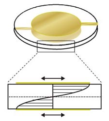 The golden standard to measure and study the cytomechanics of living cells is scanning force microscopy (SFM). It provides the stiffness of the cells with a subcellular lateral resolution so that different parts of the cells can be distinguished. We use acoustic resonators as a growth substrate for adherent animal cells and analyze their resonant oscillation with and without cells. From these readings we can also extract the micromechanics of the cells on the surface. Compared to SFM, this technique is, however, a non-imaging but integral approach. On the other hand it can be performed in a normal cell culture incubator and with a time resolution of seconds.
Besides measuring cytomechanics, we use these resonators to develop a sensor for biocompatibility screening. The resonators are coated with a thin layer of a technical material of interest and the interactions of cells with this surface are then studied by analysis of the resonant oscillation.

In Situ Electroporation - Nanoparticles
In Situ Electroporation
 The term electroporation defines the reversible permeabilization of the cellular plasma membrane by means of a short-term but strong electric field (app. 1 kV/cm). This short-term permeabilization allows those hydrophilic molecules and ions to enter the cell interior that are normally excluded from there due to their hydrophilic nature by the membrane diffusion barrier. Electroporation has been successfully applied in the past to introduce all kinds of exogeneous molecules into the cells. Of greatest importance and relevance is the introduction of recombinant DNA.
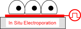
Electroporation is most often performed with cells in suspension. In situ electroporation means electroporation of cells grown on goldfilm electrodes as used in ECIS. Thus, the cells can be loaded with exogeneus molecules without being detached from their growth substrate (see image on the right). Moreover, in situ electroporation is combined with impedimetric monitoring so that the response of the cells after introducing any kind of xenobiotic can be followed online. We use this combination of in situ electroporation and ECIS to study and analyze the effects of exogeneous molecules like small organic probes, peptides, antibodies, enzymes and DNA / RNA when they become active inside the cells.
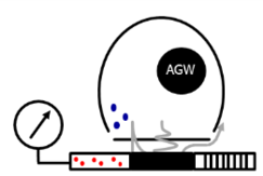
Being a member of the DFG Priority Program SPP1313 we also study the biological response of nanoparticles when they are introduced into the cytoplasm of animal or human cells by in situ electroporation.
Chemical Analysis of Animal Cells - Mass Spectrometry
Chemical Analysis of Animal Cells by Mass Spectrometry
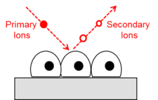
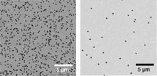
 Animal cells are chemically analyzed using a dual beam secondary ion mass spectrometer. A Bi-cluster ion source is used to generate secondary ions of the sample surface which are analyzed in a time-of-flight mass analyzer. Scanning the sample with the focused ion beam
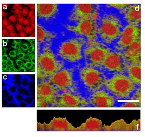
generates a 2D chemical image of the sample surface. A second ion source is used to erode the sample in between two subsequent imaging cycles. Sputter erosion removes a thin layer of the organic material ( app. 20 nm) and exposes a new, inner surface of the sample. Subsequent cycles of sputter erosion and chemical surface imaging generate a 3D image of the cell providing chemical contrast. The intensity images shown on the right are color-coded as follows: red - amino acid fragments; green: membrane fragments; blue: fragments of the growth substrate. Images a, b,c and d show the lateral distribution (xy) of the respective secondary ions in a certain depth within the sample. Image f provides a side view on the sample and, thus, distribution of chemical species perpendicular to the sample surface (xz).
This project is pursued in close cooperation with Tascon GmbH, Münster.
Barrier Analysis of 2D-Tissues - Porous Surfaces
Barrier Analysis of 2D-Tissues - Porous Surfaces
 Whole-cell biosensors based on permeable filter-like growth substrates (first image on the right) provide the option to expose the cell layer from both sides to some compound of interest. When cells are grown in these materials even the lower, substrate-facing plasma membran domain is accessible. Systems like this have been used for many years in so-called Ussing chambers to study the physiology of barrier-forming epithelial and endothelial tissues.
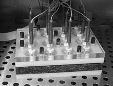
A recent prototype development from our lab (second image on the right) has provided an automated, multi-well device to perform Uss
ing-chamber like impedance measurements of epithelial and endothelial tissues to
quantify the cell layers' barrier function towards ion movement. The physical
parameter that is measured is called the transepithelial electrical resistance (TER) and
often used to describe the electrical tightness of 2D tissues. In 2008 it has been introduced into the market and
the project was awarded with the Transferpreis der University Münster as well as the Innovationspreis des Münsterlandes.
We are currently exploring several strategies to make use of these kind of porous polymer materials (lower image on the right) equipped with
different transducer functionalities on either side of the filter for novel whole-cell biosensors.

Nicole Guber - 18.03.2020 11:31