Peripheral nervous system and focal bony erosions in Rheumatoid Arthritis
Background
Rheumatoid Arthritis (RA) is a systemic chronic inflammatory condition predominantly affecting the joints and leading to destruction of articular cartilage and focal bony erosions. It has been observed that the distribution of nerve fibres in the inflamed tissue are altered from a catecholaminergic phenotype to a VIP/cholinergic phenotype but the underlying processes still remain unclear.
-->This research project focuses on the focal bony erosions in RA affected joints in the context of a possibly altered local neurotransmitter milieu and, especially on the cells responsible for bone matrix degradation, the osteoclasts. We are using an animal model of collagen-induced arthritis (CIA) to compare osteoclastogenesis at various stages of disease progression to healthy controls by gene expression analysis of typical osteoclastic markers like calcitonin receptor, cathepsin K and tartrat-resistent acid phosphatase as well as various neurotransmitter receptors for acetylcholine, vasoactive intestinal peptide and noradrenaline.
The influence of acetylcholine, VIP and noradrenaline on differentiation and activity of osteoclasts will be examined and compared between untreated animals and CIA animals.
-->![]()
![]() The first step was to establish a protocol for in vitro osteoclastogenesis from bone marrow derived macrophages. Therefore, bone marrow was harvested from femur and tibia of dark agouti rats and stimulated with M-CSF (macrophage colony stimulating factor) and the major osteoclast differentiation factor RANKL (receptor activator of nuclear factor kappa-B ligand).
The first step was to establish a protocol for in vitro osteoclastogenesis from bone marrow derived macrophages. Therefore, bone marrow was harvested from femur and tibia of dark agouti rats and stimulated with M-CSF (macrophage colony stimulating factor) and the major osteoclast differentiation factor RANKL (receptor activator of nuclear factor kappa-B ligand).
The results aim to lead to a better understanding of the communication of bone tissue and peripheral nervous system in the context of chronic inflammatory diseases like rheumatoid arthritis.
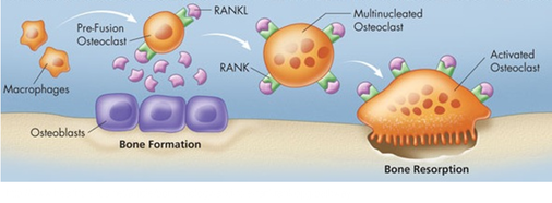
Fig. 1: In vivo osteoclastogenesis
Bone homeostasis is maintained through tightly controlled bone degradation and bone formation. Physiological bone turnover ist achieved through fine tuned activation of bone degrading osteoclasts and bone forming osteoblasts.
www.healthplexus.net/article/bone-biology-and role-rankranklopg-pathway
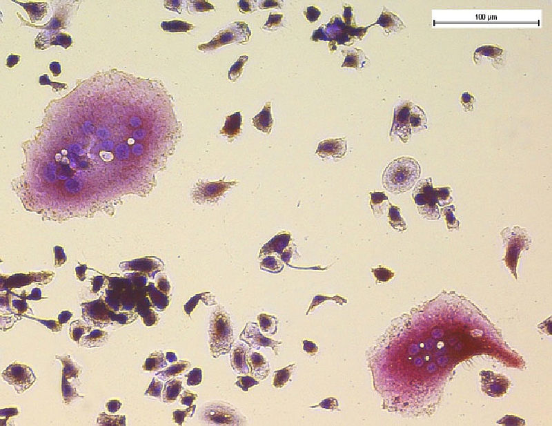
Fig 2: Osteoclast formation after 5 days of differentiation with M-CSF/Rankl. Multinucleated cells which stain positive for tartrate-resistent acid phosphatase (red) are considered as osteoclast-like cells and start to form after about 3 days of stimulation.
-->Investigators
Susanne Grässel, Prof. Dr. rer. nat.; Dominique Muschter, Dipl. Biochem.,
 Nicole Schäfer, Bachelor Biotech., PD Dr. med. Johannes Beckmann, Orthopedic Surgery, University Hospital Regensburg
Collaboration: Prof. Dr. Rainer Straub, Department of Internal Medicine I, University Hospital Regensburg
This work is supported by the Deutsche Forschungsgemeinschaft (GR 1301/10-1) as subproject of the Priority Program SPP 1468 “ImmunoBone”
-->
Susanne Graessel - 12.12.2019 11:40