

Neurobiology of Major Depressive Disorder
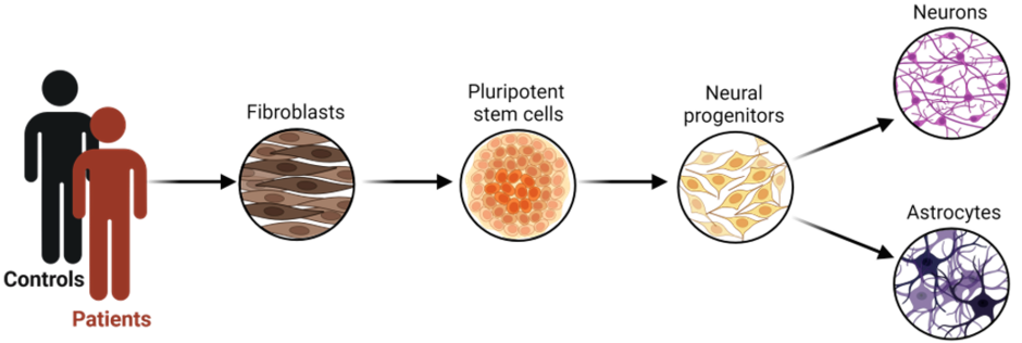
- To investigate cellular alterations in major depressive disorder (MDD), we collect skin biopsies from patients and healthy controls to culture primary fibroblasts.
We could already show that fibroblasts from MDD patients showed reduced mitochondrial function, including lower oxidative phosphorylation activity and decreased ATP levels (Kuffner et al., 2020). After reprogramming these cells into induced pluripotent stem cells (iPSCs), we differentiate them into neural progenitor cells (NPCs) which preserve this impaired mitochondrial phenotype (Triebelhorn et al., 2022). Further differentiation into neurons reveal electrophysiological changes, including lower membrane capacitance, a less hyperpolarized membrane potential, increased Na+ current density, and greater spontaneous activity (Triebelhorn et al., 2022).
We use this model to further investigate neurobiological foundations of MDD and to explore the effects of pharmacological treatments. - Chronic stress, particularly early in life, is a major risk factor for MDD and leads to structural and functional neuronal changes. Prolonged glucocorticoid exposure causes dendritic atrophy and spine loss in regions such as the prefrontal cortex and hippocampus. Cortisol binds cytosolic glucocorticoid receptors (GR, NR3C1), triggering nuclear translocation and transcriptional regulation of target genes. Ongoing work in our lab also shows rapid, direct effects of cortisol on mitochondrial function in hiPSC-derived NPCs, altering glycolysis and oxygen consumption. This indicates the involvement of a rapid, membrane-associated cortisol receptor or other non-genomic metabolic mechanisms operating alongside the genomic pathway.
- Neurons in culture self-organize into functional networks that show spontaneous, increasingly synchronized activity as synaptic connectivity develops. Using fura-2 calcium imaging, we analyze intracellular Ca²⁺ transients in 2D cultures of iPSC-derived neurons from patients with MDD and healthy controls (Issa et al., 2024). Graph-theoretical analysis reveal reduced clustering coefficient, average node degree, and global efficiency in MDD-derived networks, indicating fewer connections and less efficient functional topology at the microscale in depression.
Network topology and functional connectivity dynamics of hiPSC-derived neurons and networks will be investigated in more detail using a high-density multi-electrode array (HD-MEA).
The Enigma of the Translocator Protein 18 kDa (TSPO)
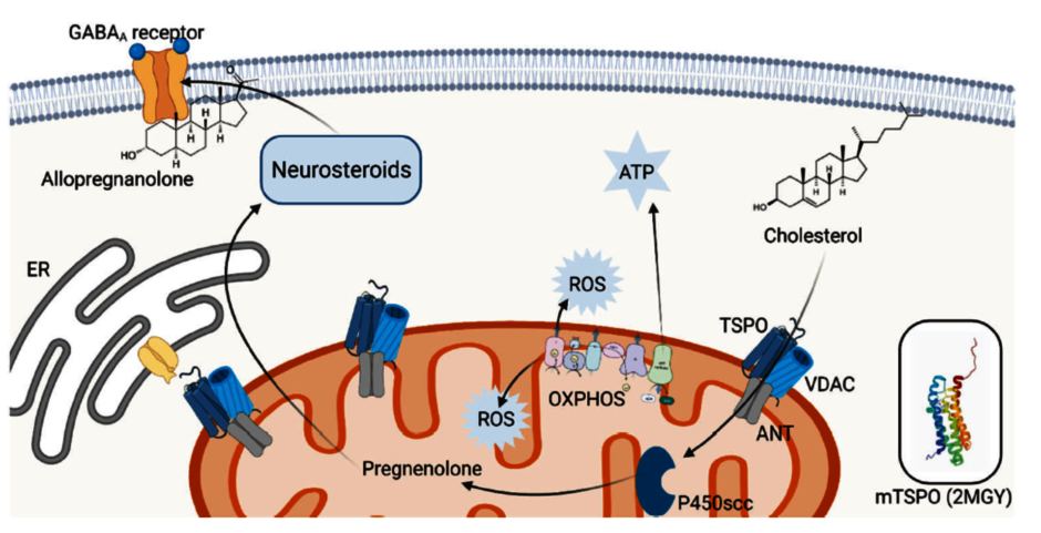
- The Translocator Protein 18 kDa (TSPO) is a highly conserved protein of the outer mitochondrial membrane with multiple important functions. We examine TSPO function in mitochondrial metabolism using CRISPR/Cas9-engineered hiPSCs lacking TSPO and matched control cells. Neural progenitor cells, astrocytes, and neurons are analyzed for bioenergetics, mitochondrial membrane potential, Ca²⁺ homeostasis, oxidative stress, and morphological mitochondrial parameters. We already showed that TSPO knockout causes mitochondrial depolarization, altered cytosolic and mitochondrial Ca²⁺ levels, increased reactive oxygen species, reduced mitochondrial DNA copy number, and smaller cell size, alongside decreased VDAC expression and respiration. Similar reductions in TSPO, VDAC1, and mitochondrial function are found in a depression cell model, linking TSPO deficiency to mitochondrial dysfunction relevant to psychiatric disease (Bader et al., 2019; 2023; 2024).
Overall, TSPO loss disrupts mitochondrial homeostasis and impairs cellular bioenergetics.
(https://www.nature.com/articles/s41380-022-01561-3 (externer Link, öffnet neues Fenster))
Methods
Cell Culture & Molecular Biology
- Reprogramming & iPSC-Technology
- Cloning, CRISPR/Cas9 Technology
Electrophysiology
- Patch-Clamp
- 2 conventional Patch-Clamp Setups
- Port-a-Patch Platform (nan]i[on)
- Orbit mini Platform (nan]i[on)
- High-Density Multielectrode Array (MaxOne, MaxWell Biosystems)
Fluorescence Microscopy
- Immunofluorescence
- Live-Cell Imaging (e.g., Ca2+ Imaging)
Mitochondrial Respirometry
- Seahorse XFp Analyzer (Agilent)